
A Trip to the European Synchrotron Research Facility
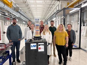
The beamtime team standing beside a stack of 3 kidneys which have been mounted for scanning.
As PhD researchers in the Tech4Health Centre for Doctoral Training, Olivia (Sheil), Isla (Henderson) and I, (Lisa Chestnutt) are carrying out our research as part of a group that uses Hierarchical Phase-Contrast Tomography (HiP-CT) to study the structure and function of human organs. HiP-CT is a high-resolution synchrotron-based imaging technique that allows the study of whole organs in remarkable detail. During our first few months of working with the research group, we had the incredible opportunity to visit the European Synchrotron Research Facility (ESRF) in Grenoble, France. HiP-CT was developed at ESRF and will play a pivotal role in our research, so we were eager to see the technique in action and gain hands-on experience with the imaging process.
Accompanied by our Professor, Peter Lee, we travelled to France and embarked on a high-throughput beamtime to image multiple hearts, lungs, and kidneys at 20-micron resolution. Scanning at the ESRF is a 24/7 operation, so we divided into three shifts. Each of us was paired with an experienced team member based at the ESRF site, who guided us through the procedures and shared their expertise of synchrotron technology. Over several days, we were immersed in the full imaging pipeline. Our responsibilities included changing samples and adjusting the sample stage to align each new organ with the beam. We also learnt to launch scans and review the data as it was produced, gaining valuable insights into how the raw data evolves into the detailed images critical to our research.
One of the highlights of the trip was visiting the LADAF anatomy facility, which supply donor organs for HiP-CT imaging. Here, we gained an understanding of the organ donation and preparation process and of the other work going on at the institution. Back at the ESRF site, we visited the sample preparation lab where we got an overview of the sample mounting process. This is an essential step in ensuring optimal image quality. Here, we learnt about the use of agar jelly to position organs securely, and the application of ethanol or formalin to fix them in place. These hands-on lessons deepened our appreciation for the hard work that goes into every scan.
While the trip was a shared experience, each of us is pursuing a distinct research focus using HiP-CT. Isla will conduct dynamic experiments to investigate the biomechanical changes in joints caused by osteoarthritis. Olivia aims to quantify the mechanical properties of blood vessel walls through dynamic synchrotron imaging. I will use HiP-CT to explore the myocardium's structure and function in a rare congenital heart defect called single ventricle disease. The trip allowed us to start planning our own experiments, informed by the insights we gained into the capabilities and limitations of the imaging process.
Amidst the busy beamtime schedule, we managed to carve out a few hours to explore Grenoble. Our climb to the top of the Bastille offered stunning views of the city and the synchrotron, and we enjoyed the chance to warm up with a hot drink as we passed through the city’s Christmas market on the way back to site.
Our visit to the ESRF gave me a deeper understanding of HiP-CT and its potential applications, and I’m grateful to the experts who took time to show us how it all works. With the new knowledge that I’ve gained, I feel better equipped to plan and conduct my own experiments using the technique. The trip was a great introduction to the possibilities of HiP-CT, and I’m excited to see how it will develop and shape our work going forward.